Scheldt species taxon details
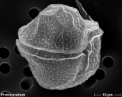
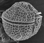
Protoceratium reticulatum (Claparède & Lachmann) Bütschli, 1885
110321 (urn:lsid:marinespecies.org:taxname:110321)
accepted
Species
marine
(of ) Claparède E. & Lachmann J. (1858). Etudes sur les Infusoires et les Rhizopodes. <em>Mém. Inst. Genev.</em> 5, 6: 489 pp. [details]
Guiry, M.D. & Guiry, G.M. (2025). AlgaeBase. World-wide electronic publication, National University of Ireland, Galway (taxonomic information republished from AlgaeBase with permission of M.D. Guiry). Protoceratium reticulatum (Claparède & Lachmann) Bütschli, 1885. Accessed through: VLIZ Consortium Scheldt Species Register at: https://www.scheldemonitor.nl/speciesregister/aphia.php?p=taxdetails&id=110321 on 2025-09-11
VLIZ Consortium. Scheldt Species Register. Protoceratium reticulatum (Claparède & Lachmann) Bütschli, 1885. Accessed at: https://scheldemonitor.org/speciesregister/aphia.php?p=taxdetails&id=110321 on 2025-09-11
Date
action
by
2006-07-27 06:59:07Z
changed
Camba Reu, Cibran
2015-06-26 12:00:51Z
changed
db_admin
original description
(of ) Claparède E. & Lachmann J. (1858). Etudes sur les Infusoires et les Rhizopodes. <em>Mém. Inst. Genev.</em> 5, 6: 489 pp. [details]
context source (Schelde) Maris, T., O. Beauchard, S. Van Damme, E. Van den Bergh, S. Wijnhoven & P. Meire. (2013). Referentiematrices en Ecotoopoppervlaktes Annex bij de Evaluatiemethodiek Schelde-estuarium Studie naar “Ecotoopoppervlaktes en intactness index”. [Reference matrices and Ecotope areas Annex to the Evaluation methodology Scheldt estuary Study on “Ecotope areas and intactness index”. <em>Monitor Taskforce Publication Series, 2013-01. NIOZ: Yerseke.</em> 35 pp. (look up in IMIS) [details]
basis of record Gómez, F. (2005). A list of free-living dinoflagellate species in the world's oceans. <em>Acta Bot. Croat.</em> 64(1): 129-212. [details]
additional source Meunier, A. (1919). Microplankton de la Mer Flamande: 3. Les Péridiniens. Mémoires du Musée Royal d'Histoire Naturelle de Belgique = Verhandelingen van het Koninklijk Natuurhistorisch Museum van België, VIII(1). Hayez, imprimeur de l'Académie royale de Belgique: Bruxelles. 111, 7 plates pp. (look up in IMIS) [details]
additional source Guiry, M.D. & Guiry, G.M. (2025). AlgaeBase. <em>World-wide electronic publication, National University of Ireland, Galway.</em> searched on YYYY-MM-DD., available online at http://www.algaebase.org [details]
additional source Tomas, C.R. (Ed.). (1997). Identifying marine phytoplankton. Academic Press: San Diego, CA [etc.] (USA). ISBN 0-12-693018-X. XV, 858 pp., available online at http://www.sciencedirect.com/science/book/9780126930184 [details]
additional source Brandt, S. (2001). Dinoflagellates, <B><I>in</I></B>: Costello, M.J. <i>et al.</i> (Ed.) (2001). <i>European register of marine species: a check-list of the marine species in Europe and a bibliography of guides to their identification. Collection Patrimoines Naturels,</i> 50: pp. 47-53 (look up in IMIS) [details]
additional source Steidinger, K. A., M. A. Faust, and D. U. Hernández-Becerril. 2009. Dinoflagellates (Dinoflagellata) of the Gulf of Mexico, Pp. 131–154 in Felder, D.L. and D.K. Camp (eds.), Gulf of Mexico–Origins, Waters, and Biota. Biodiversity. Texas A&M Press, College [details]
additional source Moestrup, Ø., Akselman, R., Cronberg, G., Elbraechter, M., Fraga, S., Halim, Y., Hansen, G., Hoppenrath, M., Larsen, J., Lundholm, N., Nguyen, L. N., Zingone, A. (Eds) (2009 onwards). IOC-UNESCO Taxonomic Reference List of Harmful Micro Algae., available online at http://www.marinespecies.org/HAB [details]
additional source Liu, J.Y. [Ruiyu] (ed.). (2008). Checklist of marine biota of China seas. <em>China Science Press.</em> 1267 pp. (look up in IMIS) [details] Available for editors
additional source Meunier, A. (1910). Microplankton des Mers de Barents et de Kara. Duc d'Orléans. Campagne arctique de 1907. Imprimerie scientifique Charles Bulens: Bruxelles, Belgium. 355 + atlas (XXXVII plates) pp. (look up in IMIS) [details]
additional source Chang, F.H.; Charleston, W.A.G.; McKenna, P.B.; Clowes, C.D.; Wilson, G.J.; Broady, P.A. (2012). Phylum Myzozoa: dinoflagellates, perkinsids, ellobiopsids, sporozoans, in: Gordon, D.P. (Ed.) (2012). New Zealand inventory of biodiversity: 3. Kingdoms Bacteria, Protozoa, Chromista, Plantae, Fungi. pp. 175-216. [details]
new combination reference Bütschli O. (1885). Dinoflagellata. In Protozoa (1880-1889). <em>Bronn‘s Klassen und Ordnungen des Tierreichs.</em> 1: 906-1029. [details]
toxicology source Satake M., MacKenzie L. & Yasumoto T. 1997. Identification of <i>Protoceratium reticulatum</i> as the biogenetic origin of yessotoxin. Nat. Toxins 5: 164-167. [details]
toxicology source Satake M., Ichimura T., Sekiguchi K., Yoshimatsu S. & Oshima Y. 1999. Confirmation of yessotoxin and 45, 46, 47-trinoryessotoxin production by <i>Protoceratium reticulatum</i> collected in Japan. Nat. Toxins 7: 147-150. [details]
toxicology source Paz, B., Riobó, P., Fernández, M. L., Fraga, S. & Franco, J. M. 2004. Production and release of yessotoxins by the dinoflagellates Protoceratium reticulatum and Lingulodinium polyedrum in culture. Toxicon 44:251-58. [details] Available for editors
ecology source Mitra, A.; Caron, D. A.; Faure, E.; Flynn, K. J.; Leles, S. G.; Hansen, P. J.; McManus, G. B.; Not, F.; Do Rosario Gomes, H.; Santoferrara, L. F.; Stoecker, D. K.; Tillmann, U. (2023). The Mixoplankton Database (MDB): Diversity of photo‐phago‐trophic plankton in form, function, and distribution across the global ocean. <em>Journal of Eukaryotic Microbiology.</em> 70(4)., available online at https://doi.org/10.1111/jeu.12972 [details]
ecology source Jacobson, D. M.; Anderson, D. M. (1996). Widespread Phagocytosis of Ciliates and Other Protists By Marine Mixotrophic and Heterotrophic Thecate Dinoflagellates. <em>Journal of Phycology.</em> 32(2): 279-285., available online at https://doi.org/10.1111/j.0022-3646.1996.00279.x [details] Available for editors
 Present
  Inaccurate
  Introduced: alien
  Containing type locality
